
Isotropic Light-Sheet Microscopy and Automated Cell Lineage Analyses to Catalogue Caenorhabditis elegans Embryogenesis with Subcellular Resolution

Examination of Mitotic and Meiotic Fission Yeast Nuclear Dynamics by Fluorescence Live-cell Microscopy

Capturing Tissue Repair in Zebrafish Larvae with Time-lapse Brightfield Stereomicroscopy

Visualization of Recombinant DNA and Protein Complexes Using Atomic Force Microscopy

Optimized Negative Staining: a High-throughput Protocol for Examining Small and Asymmetric Protein Structure by Electron Microscopy

Using Flatbed Scanners to Collect High-resolution Time-lapsed Images of the Arabidopsis Root Gravitropic Response

Microiontophoresis and Micromanipulation for Intravital Fluorescence Imaging of the Microcirculation

ZEISS Smartzoom 5: Image Acquisition How-To

Nikon N-SIM/N-STORM Super-Resolution Microscope Systems
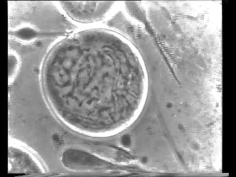
Historic time lapse movie by Dr. Kurt Michel, Carl Zeiss Jena (ca. 1943)

STED Confocal Super-Resolution - Leica TCS SP8 STED 3X

ZEISS LSM 980 - Beam Path Animation
Centrios 新成员来了!一篇了解芯片电路编辑解决方案!

Bacterial Gram Staining Procedure

Gram Staining Specimen Prep and Microscopy
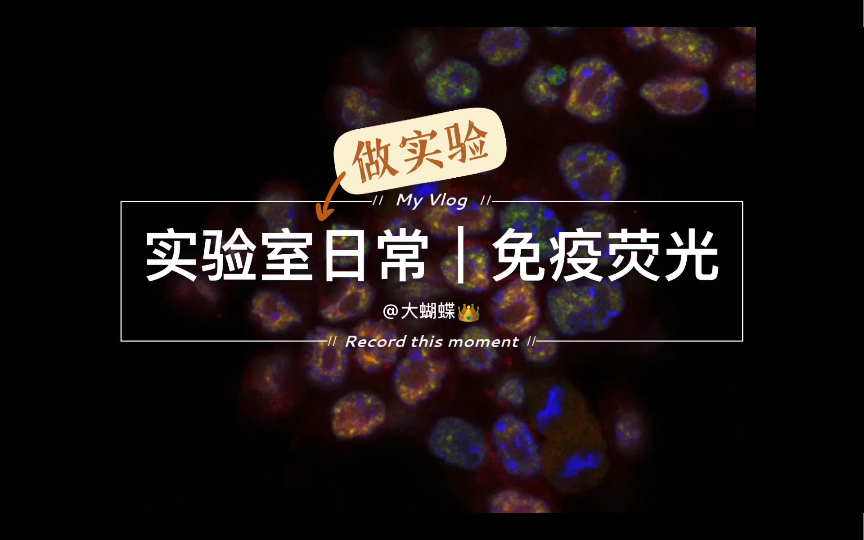
Immunofluorescence IF Protocol and Confocal Imaging

Confocal Laser Scanning Microscope Leica TCS SP8

ZEISS LSM 880 with Airyscan: Revolutionize Your Confocal Imaging

Laser Confocal Microscope | How To Set Up Confocal Multicolor Experiments

A Guide to Build a Highly Inclined Swept Tile Microscope for Extended Field-of-view Single-molecule Imaging

Intravital Microscopy of Tumor-associated Vasculature Using Advanced Dorsal Skinfold Window Chambers on Transgenic Fluorescent Mice

Detection of Microregional Hypoxia in Mouse Cerebral Cortex by Two-photon Imaging of Endogenous NADH Fluorescence

Characterization Of Multi-layered Fish Scales (Atractosteus spatula) Using Nanoindentation, X-ray CT, FTIR, and SEM

Proper Care and Cleaning of the Microscope

Single Molecule Fluorescence Microscopy on Planar Supported Bilayers

High Sensitivity Measurement of Transcription Factor-DNA Binding Affinities by Competitive Titration Using Fluorescence Microscopy

Metabolic Support of Excised, Living Brain Tissues During Magnetic Resonance Microscopy Acquisition

Intravital Imaging of the Mouse Thymus using 2-Photon Microscopy

Full-Field Optical Coherence Microscopy for Histology-Like Analysis of Stromal Features in Corneal Grafts

Live Cell Imaging of Bacillus subtilis and Streptococcus pneumoniae using Automated Time-lapse Microscopy

Direct Observation of Phagocytosis and NET-formation by Neutrophils in Infected Lungs using 2-photon Microscopy

Measuring TCR-pMHC Binding In Situ using a FRET-based Microscopy Assay

Structural Information from Single-molecule FRET Experiments Using the Fast Nano-positioning System

Automated Slide Scanning and Segmentation in Fluorescently-labeled Tissues Using a Widefield High-content Analysis System

Miniaturized Sample Preparation for Transmission Electron Microscopy

Visualizing Leukocyte Rolling and Adhesion in Angiotensin II-Infused Mice: Techniques and Pitfalls

Open Source High Content Analysis Utilizing Automated Fluorescence Lifetime Imaging Microscopy

A Novel High-resolution In vivo Imaging Technique to Study the Dynamic Response of Intracranial Structures to Tumor Growth and Therapeutics

Cell Electrofusion Visualized with Fluorescence Microscopy

Test Samples for Optimizing STORM Super-Resolution Microscopy

Analysis of Zebrafish Kidney Development with Time-lapse Imaging Using a Dissecting Microscope Equipped for Optical Sectioning

Time-lapse 3D Imaging of Phagocytosis by Mouse Macrophages

Multi-layer Cortical Ca2+ Imaging in Freely Moving Mice with Prism Probes and Miniaturized Fluorescence Microscopy

Optical Screening of Novel Bacteria-specific Probes on Ex Vivo Human Lung Tissue by Confocal Laser Endomicroscopy

Using Graphene Liquid Cell Transmission Electron Microscopy to Study in Situ Nanocrystal Etching

Horizontal Whole Mount: A Novel Processing and Imaging Protocol for Thick, Three-dimensional Tissue Cross-sections of Skin

High-resolution Episcopic Microscopy (HREM) - Simple and Robust Protocols for Processing and Visualizing Organic Materials

Intravital Microscopy of the Inguinal Lymph Node

Examining developmental processes in cancer organoids I Andreas Bausch and Anna Pastucha, TU Munich

A Simple Method for Creating a High-content Microscope for Imaging Multiplexed Tissue Microarrays

Agarose Microchambers for Long-term Calcium Imaging of Caenorhabditis elegans

Application of Membrane and Cell Wall Selective Fluorescent Dyes for Live-Cell Imaging of Filamentous Fungi
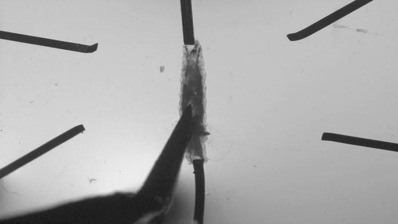
Larval body wall dissection at 3-fold time-lapse.
Separation and sorting of the ovarioles
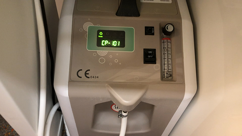
Luciferin injection and image acquisition demonstration.
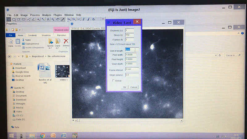
Tracking of microglial processes.
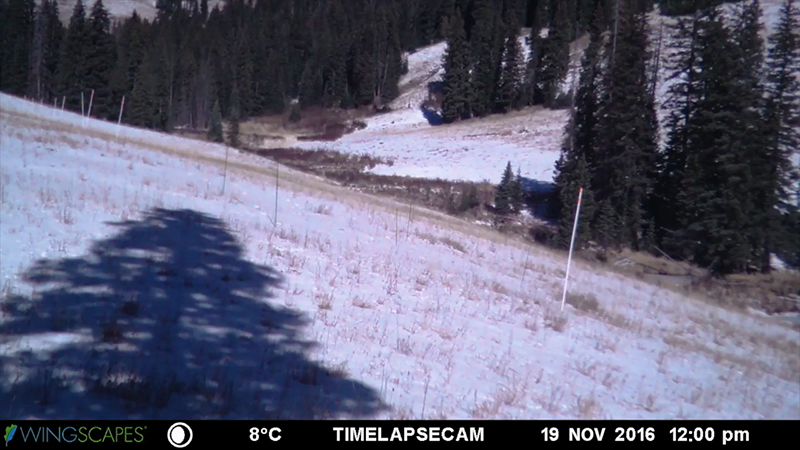
Accelerated snowmelt time-lapse during 2016-2017 snow season
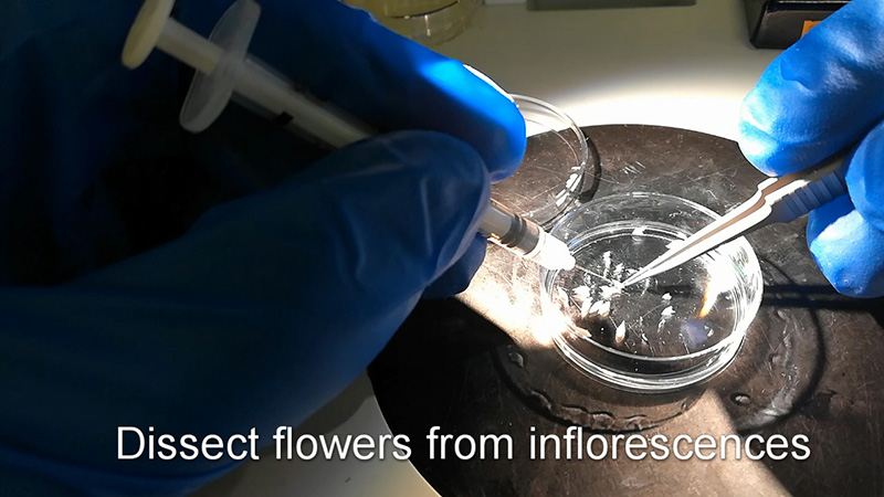
Main steps of microscope slide preparation
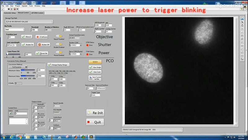
Imaging set up and real-time data acquisition of STORM imaging
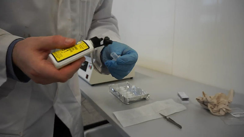
Gluing tubes onto microscope slides using UV light